
























































How Is Breast Cancer Diagnosed? Tests and Next Steps


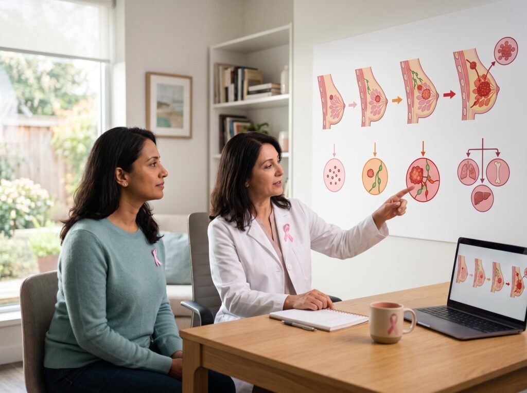
How Breast Cancer is Diagnosed usually involves more than one test. Doctors may begin with a medical history and breast examination, followed by imaging such as a diagnostic mammogram or breast ultrasound. If an area looks suspicious, a breast biopsy may be needed to determine whether cancer cells are present. Understanding the breast cancer diagnosis process can make an unfamiliar situation feel less overwhelming. A suspicious mammogram or breast lump does not automatically mean cancer. Many breast changes are caused by noncancerous conditions, and additional testing is used to determine what is actually happening.
This guide explains the main breast cancer tests, what each test can show, what happens during a breast biopsy, and what the next steps may be after a diagnosis.
What Is Breast Cancer Diagnosis?
Breast cancer diagnosis is the process doctors use to determine whether a breast abnormality or symptom is caused by cancer or another condition.
A person may enter the diagnostic process because of:
- A lump or thickened area in the breast
- A change in breast size or shape
- Skin changes
- Nipple changes or unusual discharge
- An abnormal screening mammogram
- A change noticed during a clinical breast examination
- Another breast abnormality found during imaging
The diagnostic process commonly includes a combination of physical examination, imaging, and, when necessary, tissue sampling.
Importantly, a screening test is not the same as a diagnosis. Screening is performed when someone does not have symptoms, while diagnostic testing investigates a specific symptom or abnormal finding.
How Breast Cancer is Diagnosed?
The breast cancer diagnosis process often follows several steps:
- Medical history and physical examination
- Diagnostic breast imaging
- Breast biopsy when indicated
- Pathology examination of the tissue
- Biomarker testing if cancer is confirmed
- Additional tests for staging when appropriate
Not everyone will need every test. The exact sequence depends on symptoms, imaging findings, medical history, and the clinical judgment of the healthcare team.
Step 1: Medical History and Breast Examination
The process may begin with questions about symptoms, personal medical history, family history, previous breast conditions, and other relevant risk factors.
A healthcare professional may also perform a clinical breast examination. This can involve feeling the breast and nearby areas for lumps, thickening, or other unusual changes.
A physical examination alone cannot confirm breast cancer. If something appears unusual, additional testing may be recommended.
Step 2: Mammogram and Other Imaging Tests
Imaging is an important part of many breast cancer tests. Different imaging methods provide different types of information.
Diagnostic Mammogram
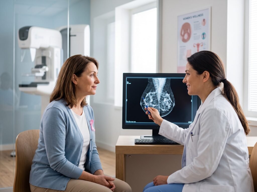
A mammogram uses low-dose X-rays to create images of breast tissue.
A screening mammogram is generally used to look for breast cancer in people without symptoms. A diagnostic mammogram is used when there is a breast symptom or when a screening mammogram has identified an area requiring closer evaluation.
Diagnostic mammography may involve additional or more detailed images of a particular area of the breast.
A mammogram may identify findings such as:
- Masses or abnormal areas
- Calcifications
- Changes in breast tissue
- Areas that require additional imaging
An abnormal mammogram does not by itself establish a cancer diagnosis. Additional imaging or a biopsy may be recommended depending on the finding.
Breast Ultrasound
Breast ultrasound uses sound waves to create images of breast tissue.
It may be used to examine an area seen on a mammogram or to evaluate a palpable lump or other breast change. Ultrasound can help distinguish between certain types of breast abnormalities, such as fluid-filled cysts and solid masses.
Ultrasound is also sometimes used to guide a needle during a breast biopsy.
Breast MRI
Breast magnetic resonance imaging, or MRI, uses a strong magnetic field and radio waves to produce detailed images.
MRI may be used in selected situations, including certain diagnostic evaluations and screening of people at high risk for breast cancer. It may provide additional information when other imaging results are unclear or when a more detailed assessment is needed.
MRI is not automatically required for every person being evaluated for breast cancer.
Understanding an Abnormal Mammogram
Receiving a report that says a mammogram is abnormal can understandably cause anxiety. However, an abnormal imaging result does not necessarily mean that breast cancer is present.
Radiologists may classify mammography findings using the Breast Imaging Reporting and Data System (BI-RADS).
Depending on the assessment, the next step may include:
- Additional imaging
- Short-term imaging follow-up
- Referral for a biopsy
- Routine screening
The appropriate follow-up depends on the specific imaging findings.
If you are asked to return for additional testing after a mammogram, follow the recommended evaluation rather than assuming the result means cancer.
Step 3: Breast Biopsy
When imaging or examination identifies an area that needs further investigation, a breast biopsy may be recommended.
A biopsy removes cells or tissue from the breast so that a pathologist can examine the sample under a microscope. A biopsy is the definitive way to determine whether an abnormal breast area contains cancer cells.
Types of Breast Biopsy
Several types of biopsy may be used.
| Type of biopsy | What it involves |
| Fine-needle aspiration | A thin needle removes fluid or cells from an area of concern |
| Core-needle biopsy | A larger hollow needle removes small tissue samples |
| Image-guided biopsy | Imaging such as ultrasound, mammography, or MRI helps guide the needle |
| Surgical biopsy | Part or all of an abnormal area is removed surgically |
Core-needle biopsy is commonly used when a tissue sample is needed, although the appropriate method depends on the characteristics and location of the abnormality.
What Happens During a Breast Biopsy?
Many needle biopsies are performed as outpatient procedures. The area may be numbed with a local anesthetic before the sample is taken.
Depending on the type of biopsy, imaging may be used to guide the needle precisely to the area of concern.
The tissue sample is then sent to a pathology laboratory for examination.
Afterward, some people experience temporary soreness, bruising, or swelling around the biopsy site. Your healthcare professional can explain what to expect based on the specific procedure.
Step 4: Understanding the Pathology Report
The pathology report is an important part of the breast cancer diagnosis process.
A pathologist examines the biopsy sample and determines whether cancer is present. If cancer is identified, the report can provide information about characteristics such as:
- The type of breast cancer
- Whether the cancer is invasive
- Tumor grade
- Other microscopic features
- Hormone receptor status
- HER2 status, when tested
These findings help the healthcare team understand the cancer and determine which additional evaluations or treatment approaches may be appropriate.
Biomarker Testing After a Breast Cancer Diagnosis
Once breast cancer has been confirmed, the cancer cells are commonly evaluated for certain biomarkers.
Two important biomarkers are:
Estrogen and Progesterone Receptors
Breast cancer cells may contain receptors for hormones such as estrogen or progesterone.
Testing for hormone receptors helps doctors classify the cancer and understand whether hormone-based treatment may be relevant.
HER2
HER2 is a protein involved in cell growth. Some breast cancers have higher-than-normal levels of HER2 or changes involving the HER2 gene.
HER2 testing helps classify the cancer and can influence treatment planning.
Other tests, including additional molecular or gene-expression tests, may be appropriate for selected patients depending on the type and stage of breast cancer.
Step 5: Breast Cancer Staging
If a biopsy confirms breast cancer, doctors may perform additional tests to determine the cancer’s stage.
Staging describes how extensive the cancer is, including whether it is limited to the breast or has involved nearby lymph nodes or other parts of the body.
Depending on the individual situation, staging may involve:
- Additional breast imaging
- Lymph node evaluation
- CT scans
- Bone scans
- PET or PET-CT scans
- Other tests selected by the healthcare team
Not every person with breast cancer needs every type of scan. The need for additional staging tests depends on factors such as the cancer’s characteristics, symptoms, and suspected extent of disease.
Breast Cancer Diagnosis: What Happens After the Tests?
The next step depends on the results.
If the imaging is reassuring
You may be advised to return to your recommended routine screening schedule or have follow-up imaging if the finding requires monitoring.
If the imaging is suspicious
Your healthcare professional may recommend a biopsy or referral to a breast specialist.
If the biopsy shows a noncancerous condition
Depending on the specific finding, you may need routine monitoring, additional evaluation, or treatment for the underlying breast condition.
If the biopsy confirms breast cancer
Your healthcare team will review the pathology results, biomarkers, and other relevant information. Additional testing may be performed to establish the stage and develop an appropriate treatment plan.
At this point, patients may be referred to specialists such as a breast surgeon, medical oncologist, radiation oncologist, or other members of a multidisciplinary cancer team.
When Should You See a Healthcare Professional?
Do not ignore a new or unusual breast change simply because it may have another explanation.
Talk with a qualified healthcare professional if you notice changes such as:
- A new breast or underarm lump
- A persistent change in breast shape or size
- Skin dimpling or unusual skin changes
- A newly inverted nipple
- Unusual nipple discharge, particularly if it is bloody
- Persistent or unexplained breast changes
Many breast symptoms are not caused by cancer, but a healthcare professional can determine whether further evaluation is appropriate.
Questions to Ask After a Breast Cancer Test
If you are undergoing breast cancer testing, it can help to write down questions before your appointment.
Consider asking:
- What did the imaging test show?
- Do I need additional imaging?
- Do you recommend a breast biopsy?
- Which type of biopsy is being considered?
- When should I expect the pathology results?
- If cancer is found, what type is it?
- Will hormone receptor or HER2 testing be performed?
- Will I need additional tests to determine the stage?
- Which specialists will be involved in my care?
Having a written list can make it easier to remember important questions during an appointment.
Frequently Asked Questions About Breast Cancer Diagnosis
1. Can a mammogram diagnose breast cancer?
A mammogram can identify suspicious breast abnormalities, but imaging alone does not always confirm cancer. If a suspicious area is found, additional imaging or a biopsy may be recommended. A biopsy provides tissue that can be examined for cancer cells.
2. Is a breast biopsy always necessary?
No. Not every breast abnormality requires a biopsy. The decision depends on the clinical examination and imaging findings. When an area is sufficiently suspicious, a biopsy may be recommended to establish whether cancer is present.
3. Does an abnormal mammogram mean I have breast cancer?
No. An abnormal mammogram means that the radiologist has identified a finding that may require additional evaluation. Many abnormal or recalled mammograms do not ultimately result in a breast cancer diagnosis.
4. How long does a breast biopsy take?
The time depends on the type of biopsy and how it is performed. Many needle biopsies are outpatient procedures. Your healthcare professional can explain the expected duration and recovery for the specific procedure being recommended.
5. What happens if my biopsy confirms breast cancer?
Your healthcare team will review the pathology report and may perform biomarker testing and additional evaluations. These results help determine the type and stage of breast cancer and guide treatment planning.
6. What is the difference between screening and diagnostic mammography?
Screening mammography is generally performed when there are no breast symptoms. Diagnostic mammography is used to evaluate a specific symptom or an abnormality identified during screening.
7. Who should I talk to about an abnormal breast test?
Start with your healthcare professional or the clinician who ordered the test. Depending on the findings, you may be referred to a breast specialist, radiologist, surgeon, oncologist, or another appropriate healthcare professional.
Conclusion
Understanding breast cancer diagnosis can make the testing process easier to navigate. A typical evaluation may involve a clinical examination, mammogram or other imaging, followed by a breast biopsy when an abnormal area needs to be examined more closely. A suspicious imaging result does not automatically mean cancer. The biopsy and pathology evaluation are central to confirming a diagnosis, while biomarker testing and staging help doctors understand the cancer more completely after it has been confirmed.
If you notice a new breast change or receive an abnormal breast imaging result, speak with a qualified healthcare professional and follow the recommended next steps. Early evaluation can help ensure that concerning changes are investigated appropriately.